7. Imaging the vertebral arteries
Imaging of the vertebral arteries is part of a standard extra-cranial cerebrovascular ultrasound duplex scan. The vertebral arteries are smaller, located more distal from the transducer and partially “hidden” behind the bony structures of the vertebrae. Therefore they are more difficult to image. Despite these limitations it is possible to visualize and obtain flow information in the vertebral arteries in the vast majority of patients. To achieve optimal results it is important to understand the topography of the vertebral arteries and to use all possible ultrasound modalities (B Mode, color Doppler and spectral Doppler) and practice. It is important to recognize that Magnetic Resonance Angiography, CT Angiography and Digital Subtraction Angiography are superior (sensitive) to ultrasound in the detection of vertebral artery pathologies. In contrast extra-cranial ultrasound has numerous advantages:
- It is less complex
- It carries no risk
- It is more cost effective
- Can be applied bedside
- More available
- Provides hemodynamic information
7.1 What is the anatomy of the vertebral arteries
The vertebral arteries arise bilaterally from the subclavian artery they travel posterior to the carotid artery (in the back of the neck) cranially to enter the skull through the vertebral canal. Both vertebral arteries join to form the basilar artery.
Note: The left vertebral artery arises from the aorta in approximately 3-5% of patients
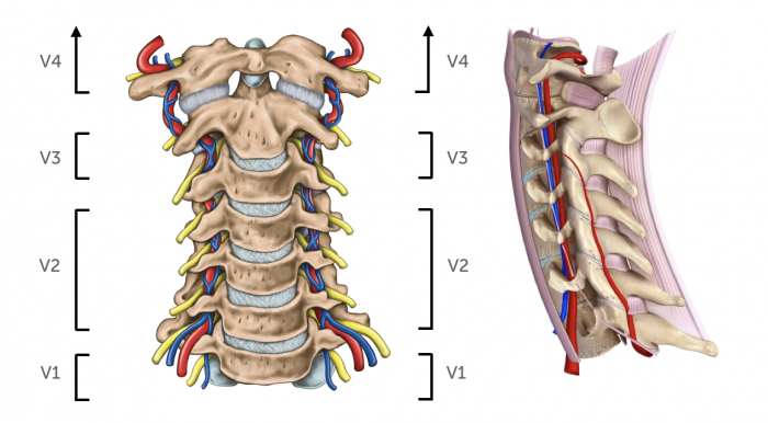
| Vertebral arteries - Topography | ||
|---|---|---|
| Segments | ||
| Origin | Subclavian artery | Origin is visible in 60-70% |
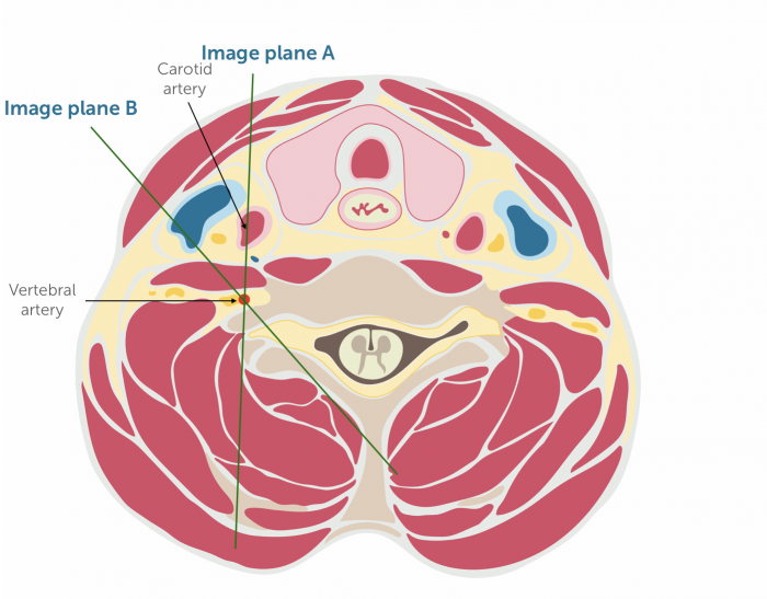
| Location | The vertebral arteries are posterior and lateral to common carotid artery | Can be imaged behind the carotid artery or from a more laterally angulated position (or by moving the transducer more lateral) |
| Course | Runs in the transverse foramen of the cervical vertebra. Enters the skull through the foramen magnum | Bony structures obscure the view. The vessels is interrupted on the US image. Tortuosity can be found (especially V1) |
7.2 Which regions do the vertebral arteries supply?
The vertebral arteries supply blood to the cranial part of the spinal cord, the brainstem and the cerebellum as wall as the posterior part of the brain. A stroke (occlusion, embolizaton) of the vertebral artery will affect the posterior circulation of the brain. Approximately 20-25% of all strokes affect the vertebrobasilar system. Strokes of the large arteries of the posterior circulation will often be fatal or cause severe disability. Typical symptoms include: ataxia vertigo, nausea, slurred speech or double vision. The vertebral arteries also play an important role as a collateral pathway in the setting of a proximal subclavian artery occlusion / stenosis by providing blood to the arm (subclavian steal syndrome). The vertebral arteries / basilar artery as part of the circle of Willis can also provide collateral blood flow in the setting of carotid artery occlusion. Conversely, occlusion of the vertebral artery does not invariable cause a stroke. Possibly the vertebral circulation is also protected by collateral blood flow through ascending cervical branches.

7.3 How can you find the vertebral arteries with ultrasound?
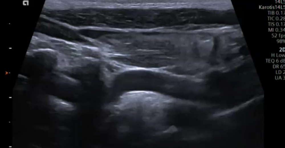
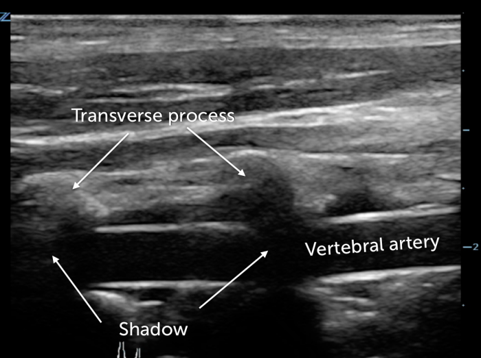
It can be challenging to visualize the vertebral arteries. Especially if they are small (hypoplastic) or occluded (no flow signal). Imaging of the vertebral arteries is more difficult in patients with “thick” short necks / obese patients. The vertebral artery is best visualized in a longitudinal view. Start by imaging the middle portion of the vertebral artery (V2) and then move caudally (to display the origin of the vertebral artery) and then more cranial to image the more distal portions of the vessel.
- Display the common carotid artery in a longitudinal view
- Slide laterally more posterior
- Angulate more posterior
- Display the vertebrae with the transverse processes (characterized by intermittent areas of shadowing) in between you will find the vertebral artery
Alternatively one can also use a more anterior transducer position imaging the vertebral arteries behind the common carotid artery.
7.4 What is the normal size of vertebral arteries?
| Normal size | 3-5mm |
| Hypoplasia | < 2-3mm or side difference of > 1:1.7 (note in hypoplasia one can often find a larger vertebral artery on the contralateral side) |
| Left vs. right | The left is more commonly larger than the right |
| Aplasia | No vertebral artery present - uncommon - 1% of population |
| Impact | The posterior circulation is more vulnerable to ischemia in patients with vertebral artery hypoplasia |
7.5 How do I use color Doppler in the assessment of the vertebral arteries?
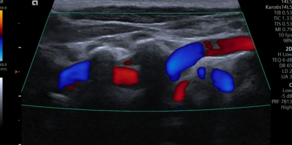
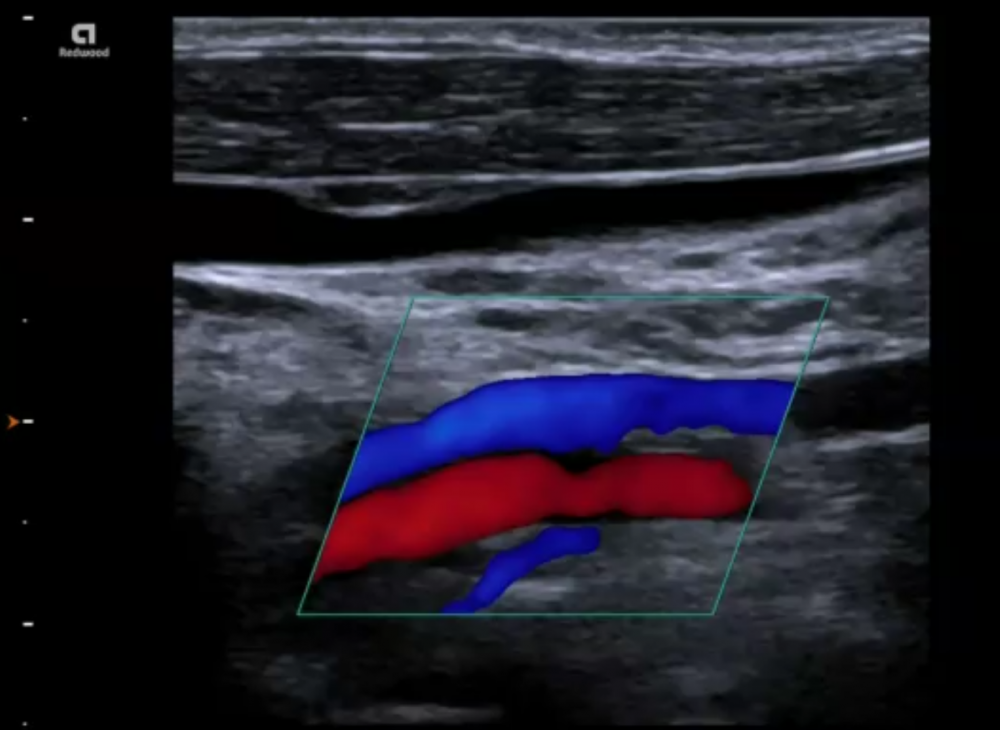
Color Doppler is helpful to locate the vertebral artery. Make sure that you optimize the color Doppler box to the direction of flow. Color Doppler is also used to determine the direction of flow and to differentiate the artery from the vein. Areas of turbulent flow indicates a stenosis. The absence of a color flow signal within the vertebral artery indicates occlusion of the vertebral artery.
7.6 What is the role of spectral Doppler of the vertebral artery?
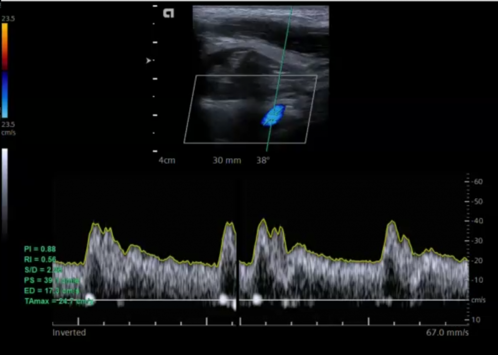
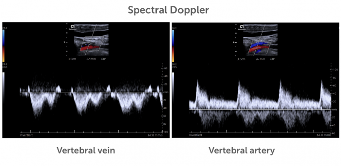
Spectral Doppler should routinely be performed during an ultrasound exam of the extracranial arteries. It allows you to:
- Differentiate the vertebral artery from the vertebral vein
- Determine the direction of flow
- Demonstrate flow within the vertebral artery
- Help to detect a vertebral artery stenosis
- Assess a dissection of the vertebral artery.
7.7 How can I determine the direction of flow in the vertebral artery?
In the setting of a subclavian steal syndrome (occlusion of the proximal subclavian artery you will find a reversal of flow in the vertebral artery). In other words the detection of a reversal in flow will allow you to diagnose /suspect an occlusion of the subclavian artery stenosis. There are several ways how ultrasound can be used to determine the direction of flow
| How to determine the direction of flow in the vertebral artery | |
|---|---|
| Color bar | Use the color bar and the vessel orientation / flow to determine the direction of flow (see below) |
| Compare CCA | Compare the flow direction (color) with that of the CCA (displayed on the same image). Both should show the same direction of flow |
| Compare with vertebral veins | Compare the flow direction (color) with that of the vertebral veins |
| PW Doppler | Use PW Doppler for the direction of flow |
If you like the way we teach, please leave a message!

