3.5.2 Size of the right atrium and associated structures
The right atrium is usually slightly smaller than the left one, and is largest in the superior inferior extension. To a certain degree the size and shape of the right atrium is also influenced by the dimensions of the left atrium. An enlarged left atrium will "stretch" the right atrium. The latter will then appear more oval in shape. Enlargement of the right atrium may occur alone or may be combined with enlargement of the left atrium. In any case, right atrial enlargement points to a disease.
Causes of RA dilatation:
- RV failure
- Tricuspid regurgitation
- Tricuspid stenosis
- Atrial septal defects
- Atrial fibrillation
- Restrictive cardiomyopathy
- Pulmonary hypertension
- Congenital abnormalities
Right atrial enlargement is a marker of the severity of disease, and also predicts outcome in some situations. These include tricuspid regurgitation, pulmonary hypertension, and right heart failure. Enlargement of the right atrium may result from right atrial volume or pressure load. In this setting you will also see a dilated inferior vena cava and bulging of the interatrial septum to the left.
Right atrial enlargement and bulging of the interatrial septum to the left in a patient with severe tricuspid regurgitation:
The size of the right atrium is measured by the same methods as those used to measure the left atrium. Thus, both distance and volume measurements can be obtained. In any case, perform at least one measurement to determine the size of the right atrium, such as the long-axis dimension of the right atrium on a four-chamber view. This parameter is easy to obtain and provides a good estimate of right atrial size. The upper limit of normal for the long axis dimension of the right atrium is 45 mm.
| Normal (mm) | 29—45 |
|---|---|
| Mild (mm) | 46—49 |
| Moderate (mm) | 50—54 |
| Severe (mm) | ≥ 55 |
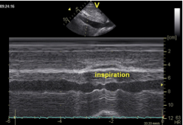
The size of the inferior vena cava provides valuable information. It permits estimation of right atrial pressure and assessment of fluid status. If the vena cava is dilated you will frequently observe dilatation of the hepatic veins. The size of the inferior vena cava is best measured from a subcostal window shortly before it enters the right atrium. The normal size of the inferior vena cava is 15 to 17 mm, with a more than 50% size reduction during inspiration. Fluctuations in size can be assessed with 2D imaging and the MMode.
Inferior vena cava; normal:
Inferior vena cava; severely dilated:

Do not measure the inferior vena cava at the site where it passes through the pericardium. There is usually some degree of narrowing there.
The size of the vena cava inferior reflects the patient's fluid status and central venous pressure, and permits estimation of RA pressure.
In athletes the inferior vena cava is often larger despite normal right atrial pressure.
Vena Cava Inferior
Size < 17mm, Inspiratory collapse ≥ 50%
The ostium of the normal coronary sinus is 12 ±2mm. A diameter above 15 mm should cause the investigator to suspect a persistent superior left vena cava. This is a congenital abnormality in which a second left-sided superior vein (embryonic remnant) drains blood from the upper left quadrant of the body via the coronary sinus into the right atrium. Other possible causes of coronary sinus enlargement are:
- Elevated RA pressure
- Vena cava sin. persistens
- Malformation (Aneurysm/Diverticula)
- Unroofed coronary sinus
Coronary Sinus
Reference Value = 4-8mm (upper limit 15m)