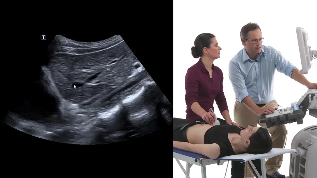
Abdominal Ultrasound BachelorClass
Watch free lectures
Create a free account or log in to access free lectures for this course.
No payment required.
Create free account Log in


What you get with this course
Curriculum
Watch free lectures
Create a free account or log in to access free lectures for this course.
No payment required.
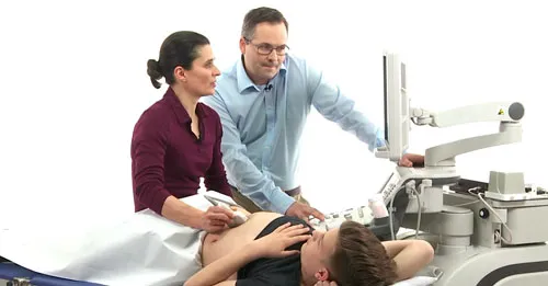
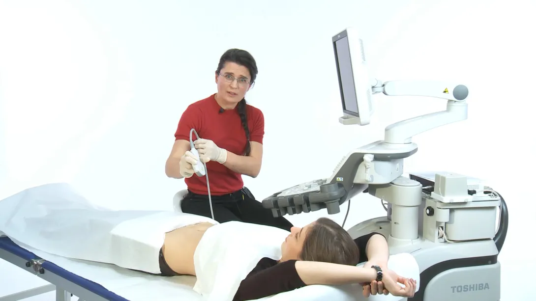
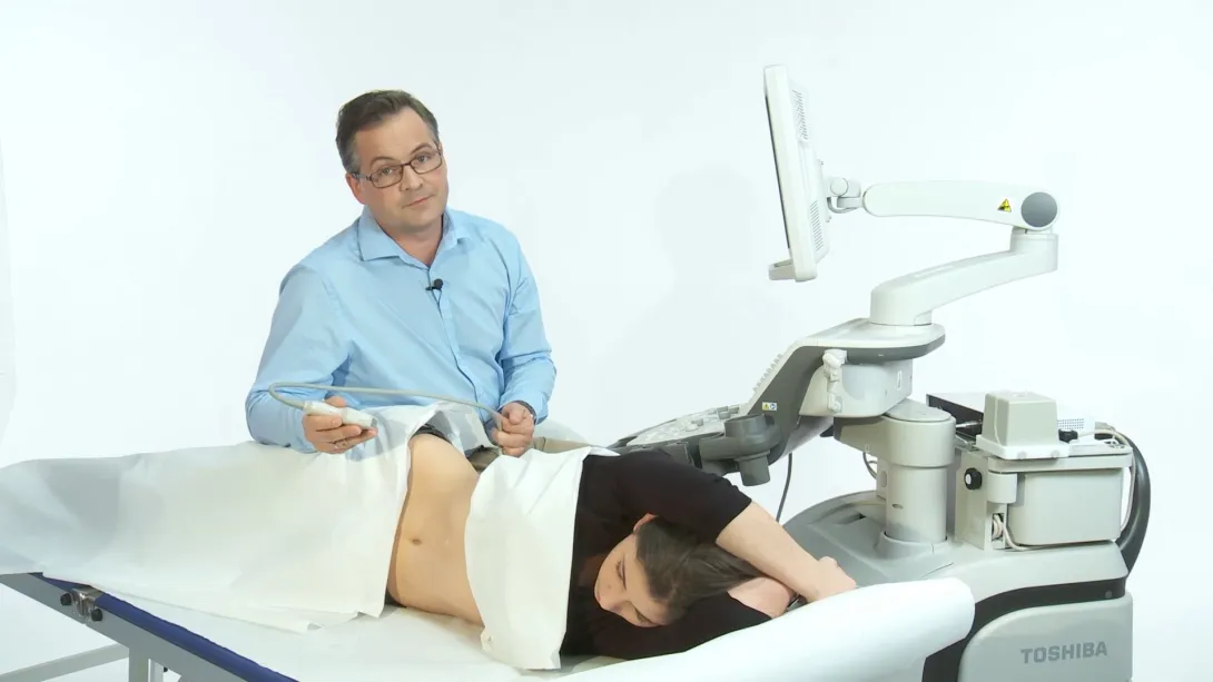
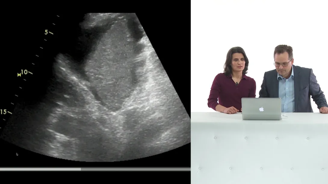
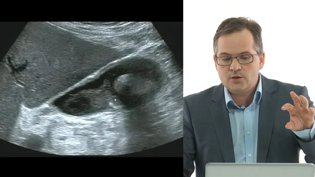
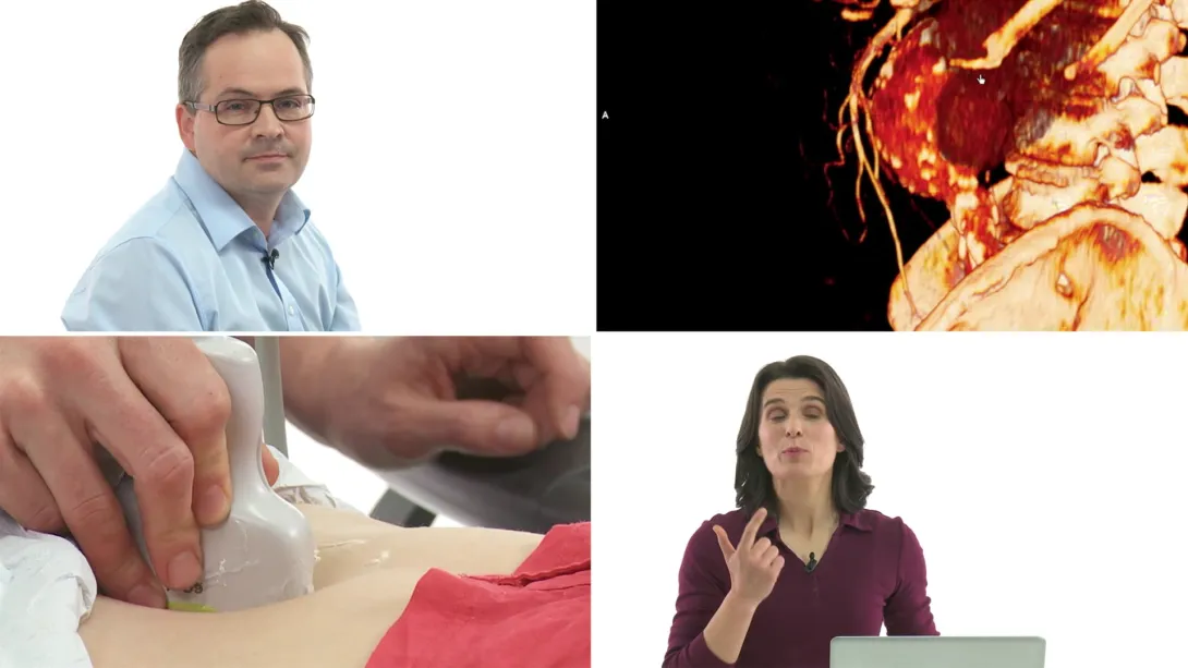
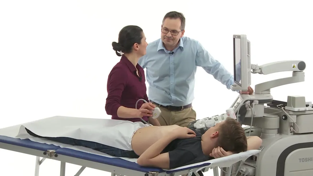
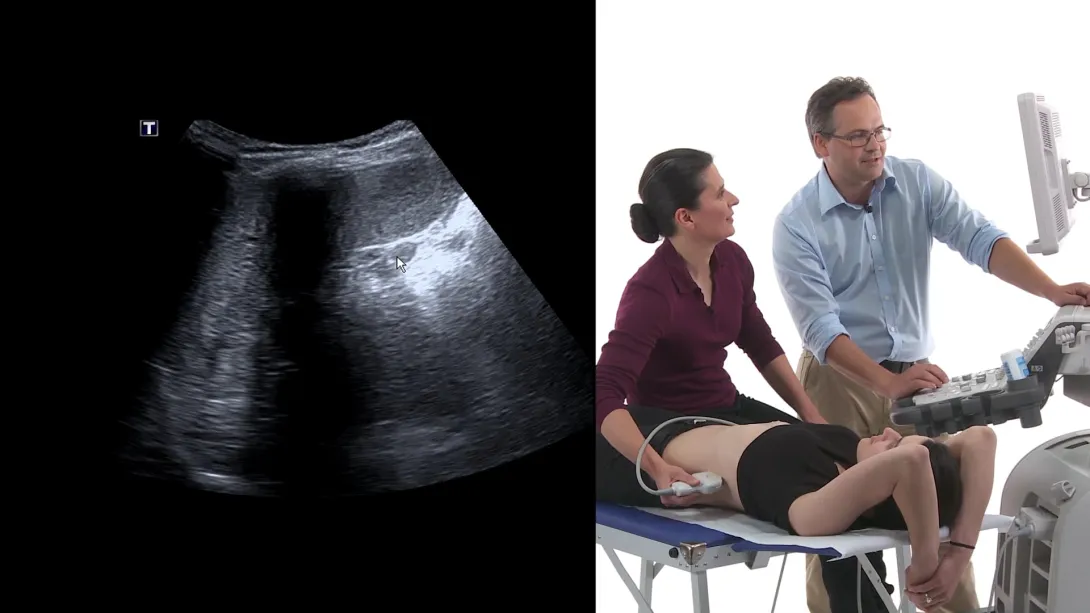
Create free account Log inYour starting point into the world of abdominal ultrasound. Meet your teachers Ulrike and Christian. Six different case reports from our lab that will show you how powerful abdominal ultrasound is. Learn how to perform an ultrasound scan on the urinary bladder and the gallbladder.
Lectures & Quizzes:
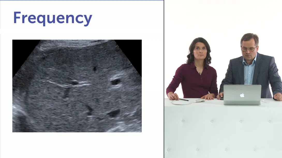
Meet the star of this course: the ultrasound wave! Principles of scanning. How to obtain really good images. How to optimize your settings and adjustments on the machine. Safety aspects. Understand the most important ultrasound artifacts and use them to your advantage.
Lectures & Quizzes:
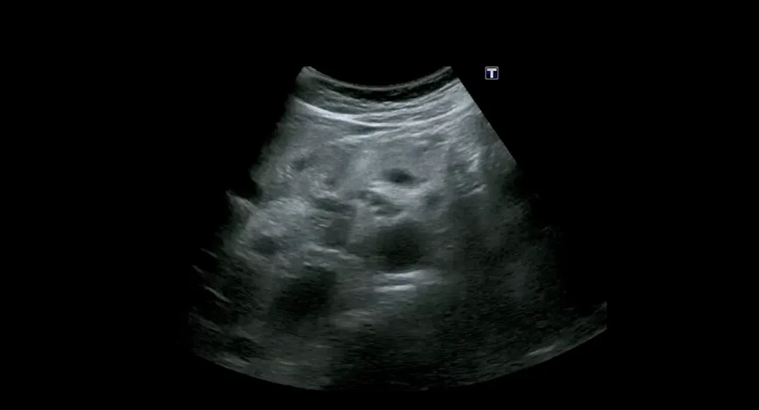
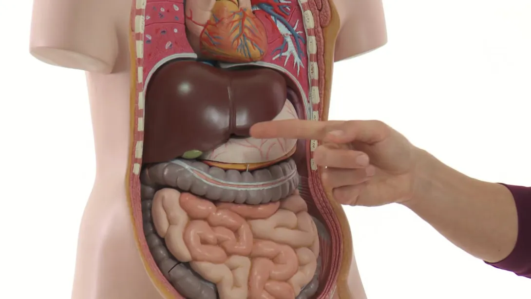
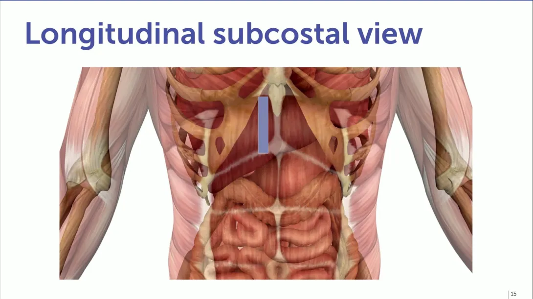
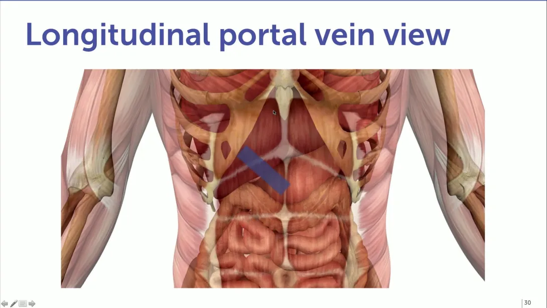
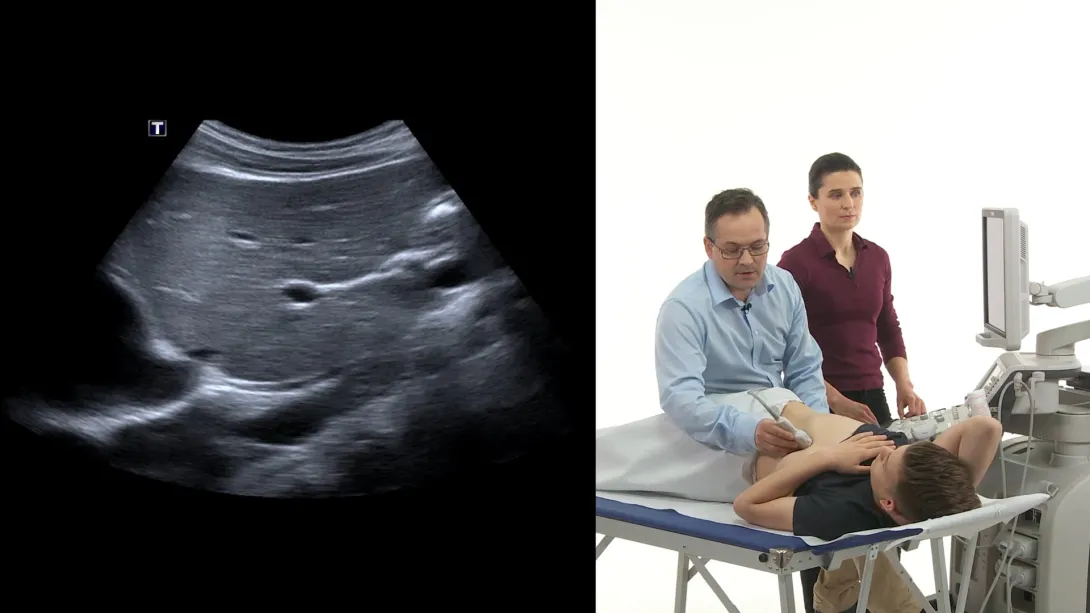
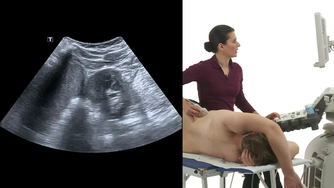
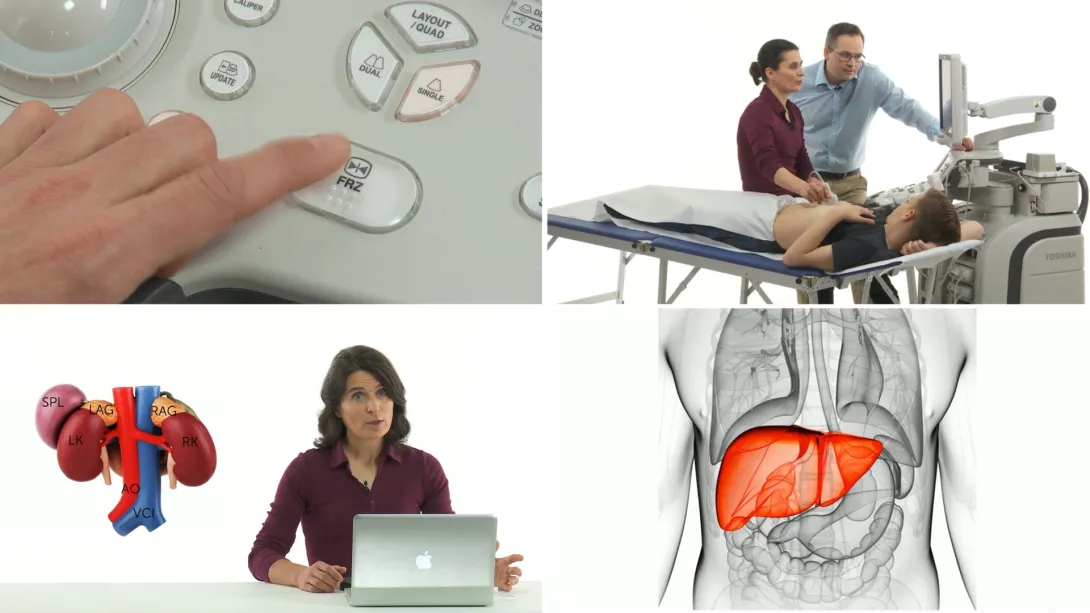
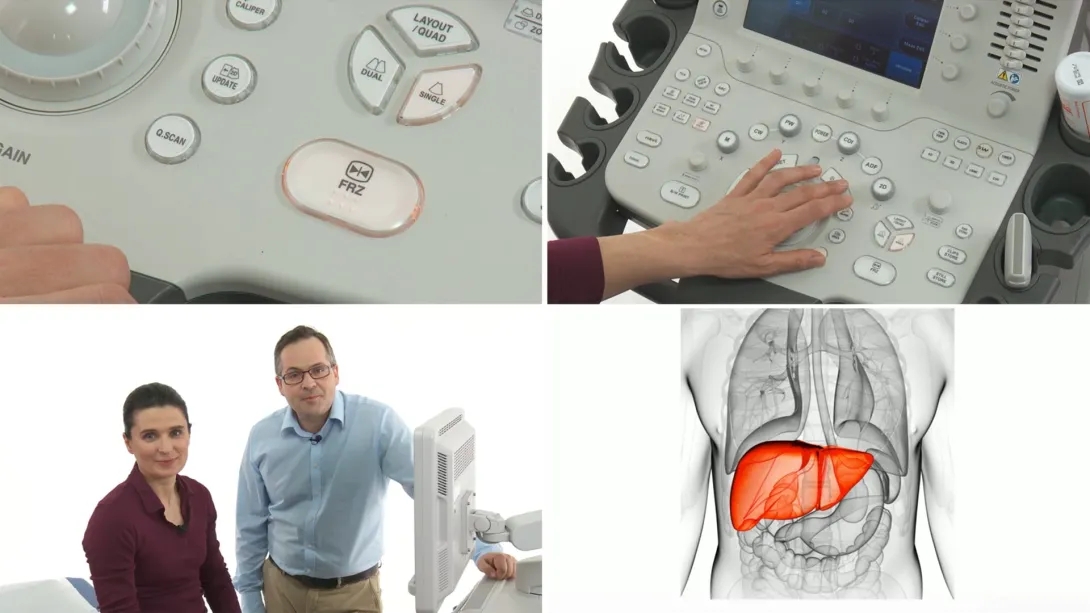
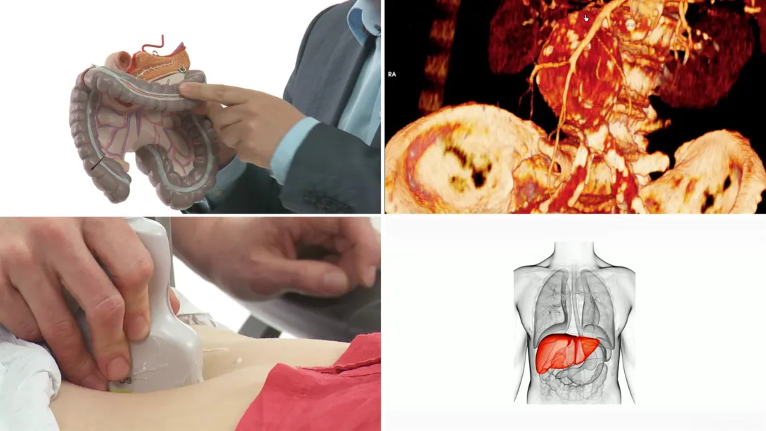
The liver is the second largest organ in our body. We start with a review of the relevant anatomic features and the important structures surrounding the liver - we will show you how to scan the liver in the standard imaging planes systematically, including the “emergency view” of the liver. Also see the course regarding how to perform a thorough liver scan.
Lectures & Quizzes:
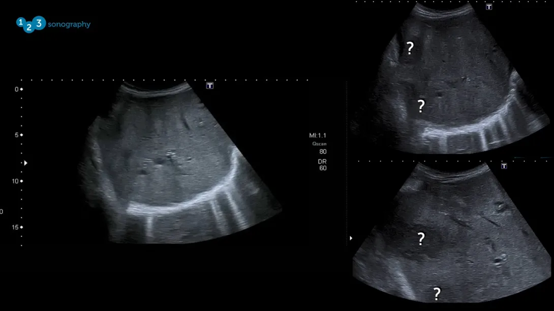
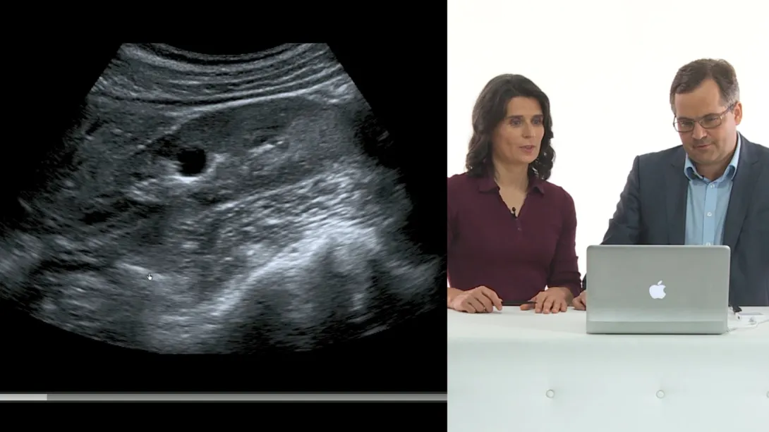
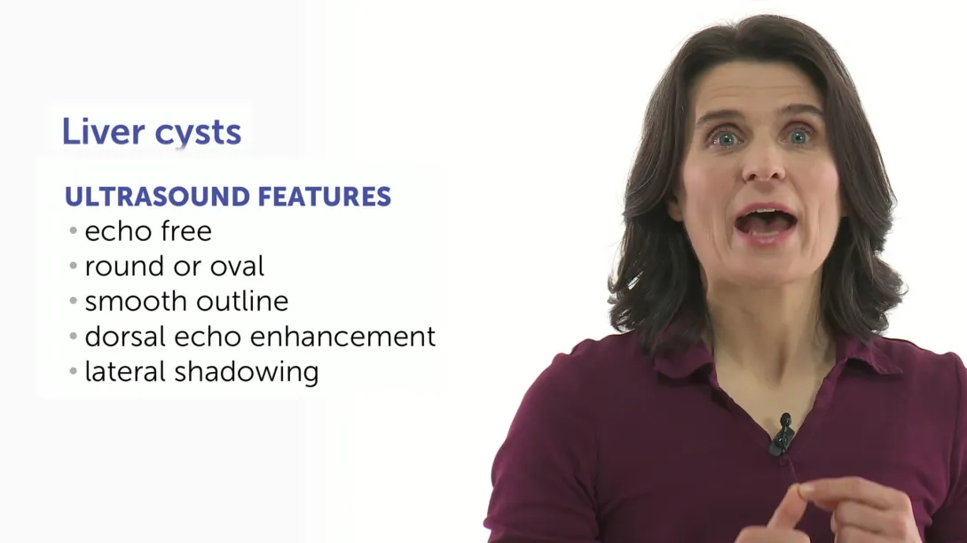
Learn how to detect and distinguish diffuse liver diseases like liver congestion, steatosis and liver cirrhosis as well as focal liver lesions with your magic ultrasound wand - we’ll take you there! What does a snowball have in common with liver lesions? You’ll find out here. After completing this chapter, you will definitely be able to differentiate between the most common liver pathologies in ultrasound. Three case reports included - test your knowledge!
Lectures & Quizzes:
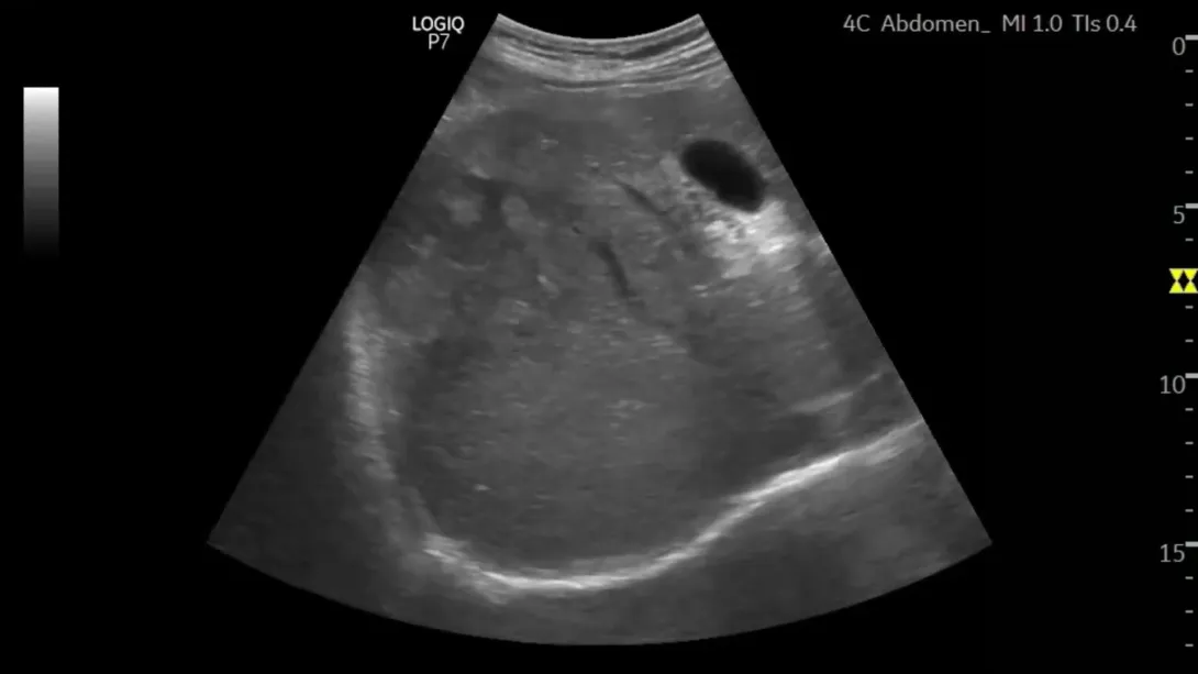
How can I detect gallstones with ultrasound? What are the criteria for cholecystitis? What are the sonographic signs for cholestasis? In this chapter we will teach you not only how to successfully find and scan the gallbladder but also how to detect and distinguish the most important pathologies of the gallbladder and the bile ducts.
Lectures & Quizzes:
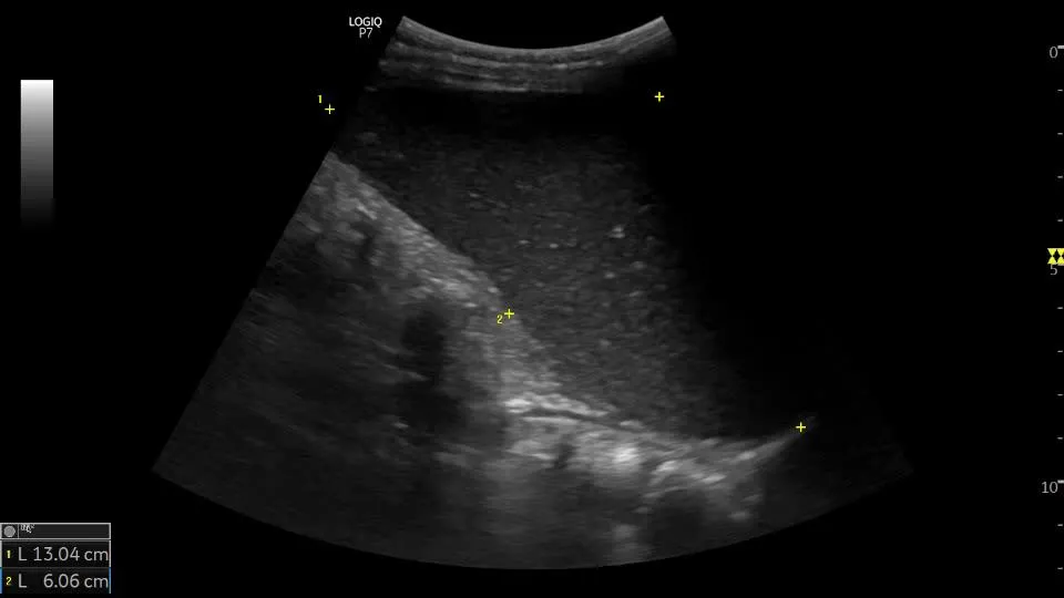
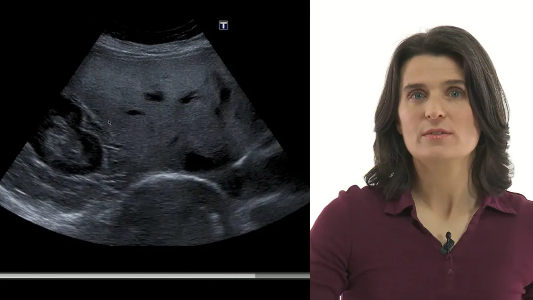
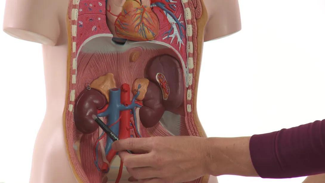
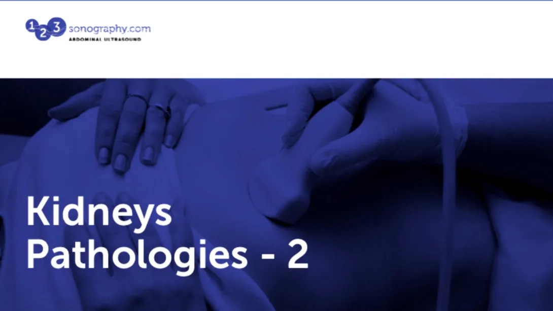
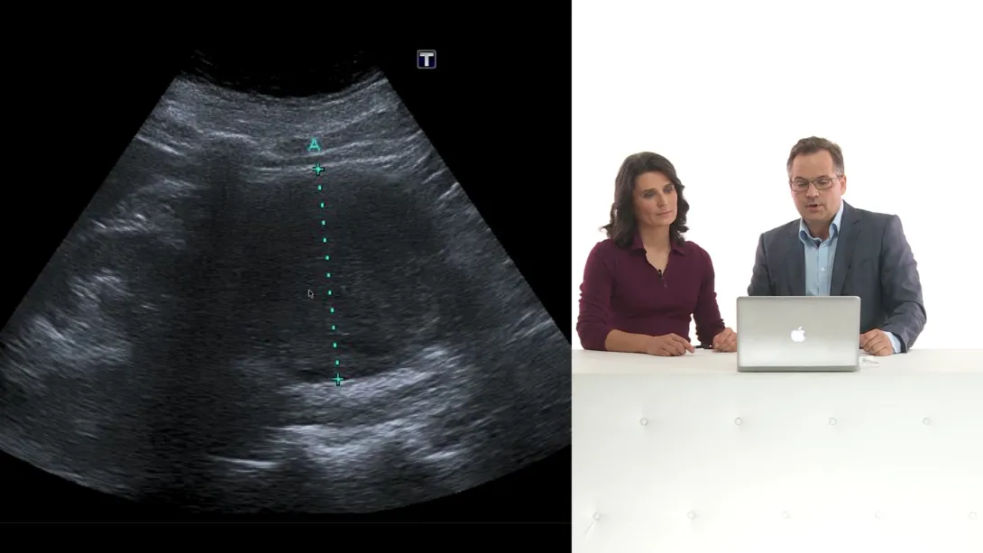
We’ll start with a review of the anatomic features and go on with the different approaches and scan planes for both kidneys - getting you ready for evaluating renal pathologies like hydronephrosis, urinary tract stones, renal cysts and tumors and the atrophic kidney disease. You will see how the kidneys can vary in size, form and position - we will make sure, you’ll always find them. Test your knowledge in a clinical context and see if you can solve a few cases.
Lectures & Quizzes:
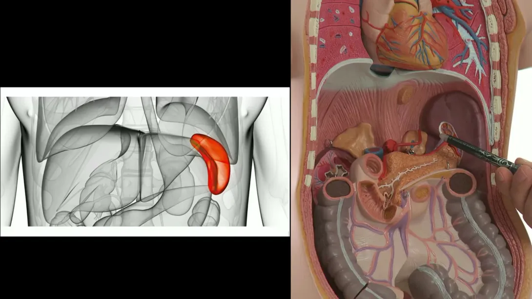
Scanning and evaluating the spleen and the rather tricky region of the adrenal glands with ultrasound will be no big deal for you after you have completed this chapter! Do you know which pathologies you should look for? Why they are so important to find? These are the topics you will learn here.
Lectures & Quizzes:
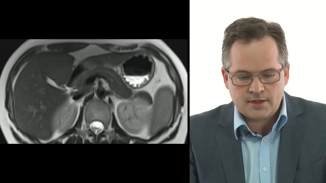
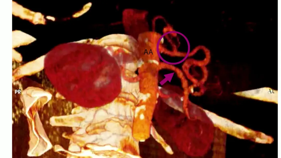
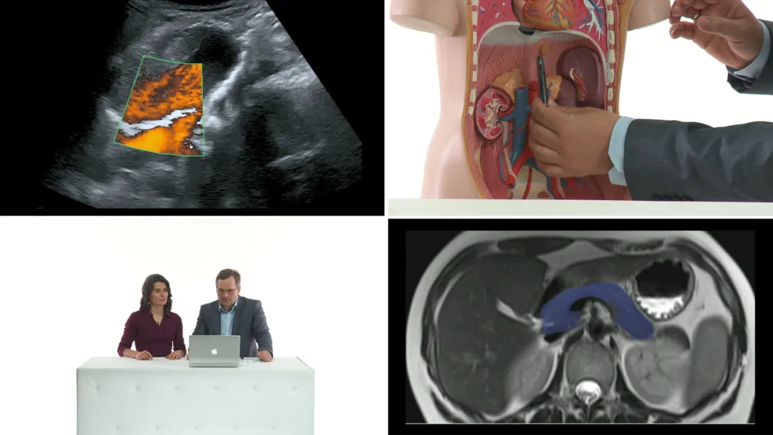
Pancreas - is also called the hidden organ? True, it is difficult to find. But not for you: once you have completed this chapter, you will never fail to find the pancreas again. We will deal with the most common pathologies of the pancreas and get on with scanning the region of the great abdominal vessels. Abdominal aortic aneurysm - early diagnosis can save lives! Be prepared for critical care scenarios and emergencies and learn how to scan and evaluate the abdominal aorta.
Lectures & Quizzes:
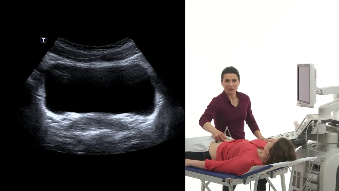
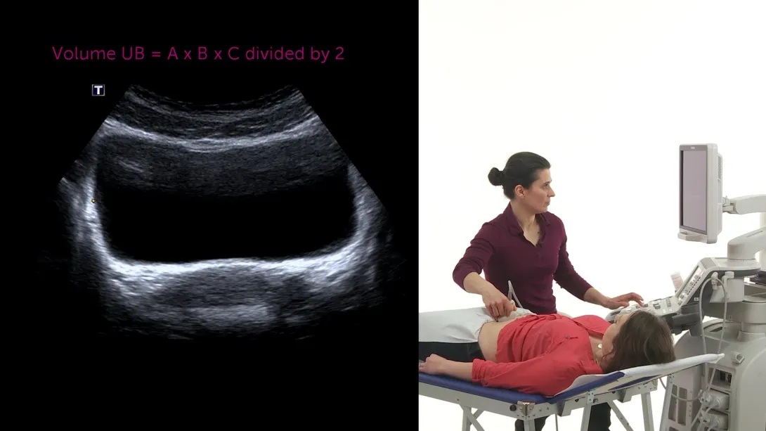
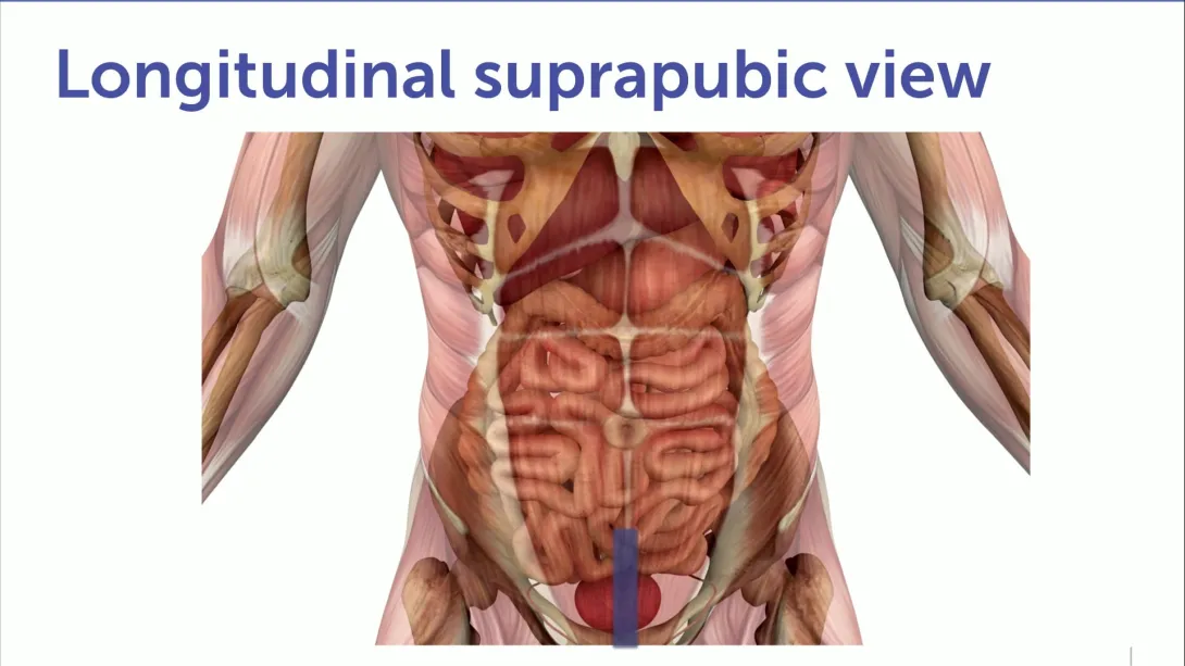
Scanning the urinary bladder with ultrasound is easy and rewarding - you’ll understand why in this lecture! Urinary retention - a painful but often easily solved problem when detected with ultrasound - you can do it! This chapter also covers the issue of residual urine and the ultrasound appearance of bladder tumors.
Lectures & Quizzes:
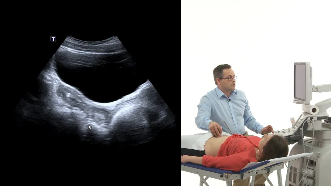
This chapter covers the different scan planes for examining the female and male pelvis: how to find the uterus, the ovaries or the prostate. Tumours and cysts and free fluid in the female lower abdomen? Prosthetic hyperplasia? After completing this chapter, you will definitely be able to detect these pathologies! We will also deal with another very important issue: the carcinoma of the prostate.
Lectures & Quizzes:
Scanning only - a complete review of all scan planes and approaches to: the liver, the kidneys and the adrenal glands region.
Lectures & Quizzes:
In this chapter you'll get the final touch - recapitulating all scan planes and approaches for spleen, pancreas and great abdominal vessels followed by scanning the urinary bladder as well as the female and male lower abdomen. Then the grand finale: we´ll demonstrate a complete abdominal ultrasound examination. Now you’re ready!
Lectures & Quizzes:
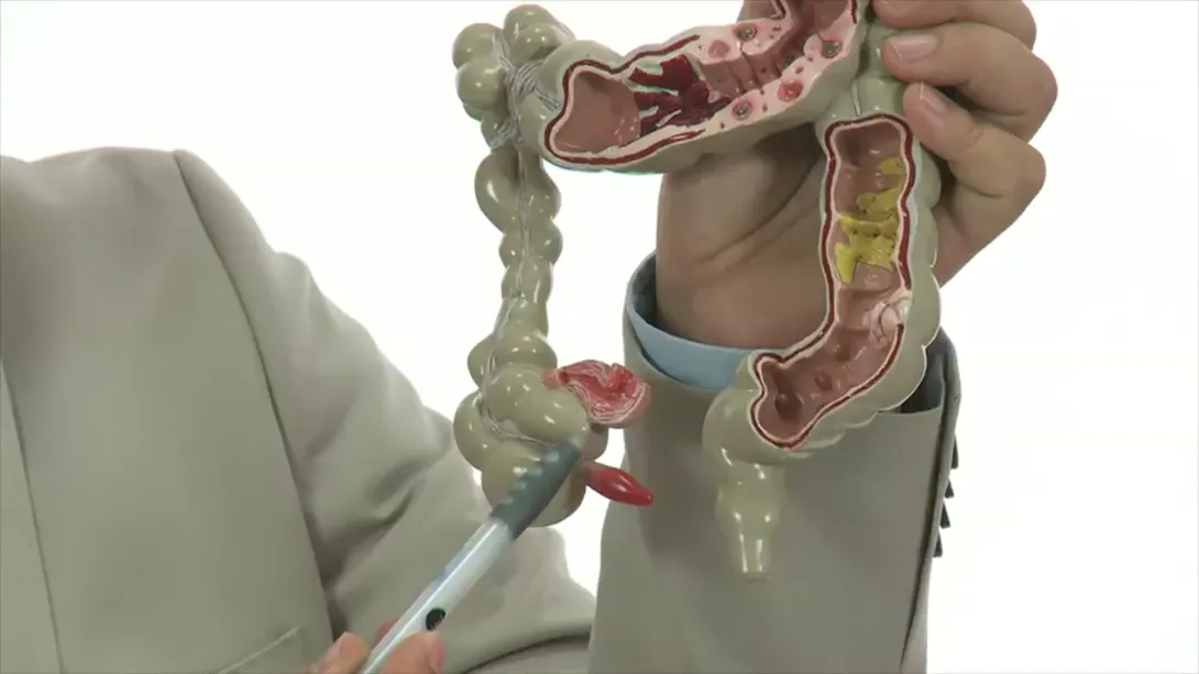
In this chapter, we will show a simple, workable approach to ultrasound imaging of appendicitis - suitable for anybody who is willing to learn abdominal ultrasound. Consider a scenario in which one of your patients is complaining about pain in the right lower quadrant of the abdomen, and has not had an appendectomy. In the case of appendicitis, an acute operation is necessary, so it is extremely important to get the right diagnosis very quickly. Appendicitis (acute inflammation of the appendix) is consistently one of the most critical differential diagnoses. In most cases, it is possible either to image the inflamed appendix with ultrasound or at a minimum to locate some characteristic ultrasound features that point to appendicitis. This premium chapter will cover everything you need to know, in order to help your patients in this emergency situation.
Lectures & Quizzes:
Learn from leading experts


Ideal for
Objectives
After completing the Abdominal BachelorClass, you will be able to perform a standard exam.
You will know the limitations of this technique.
And be able to identify the most common pathologies of all major abdominal and pelvic organ systems.

Student Discount
Are you a student? Get 50% discount on this course by completing the student application form.
Get Student DiscountPricing
Monthly Subscription
One plan. Full flexibility.
Take the most flexible route with a monthly subscription.
You get:
- Cancellation possible anytime after 4 months minimum run time
- Ability to complete quizzes and earn CME credits

Prefer to pay once?
Save more with our one-time payment options.
Half-Year Access
Our shortest option for very fast learners. Ideal for people who have plenty of time to learn.
You get:
- 6 months access to our course
- Ability to complete quizzes and earn CME credits
One-Year Access
Our most recommended access duration to dive deep into the course. Save 30% on the 6-month option.
You get:
- 12 months access to our course
- Ability to complete quizzes and earn CME credits
Two-Year Access
If you want to take your time learning, this option is perfect for you. Save 40% on the 6-month option.
You get:
- 24 months access to our course
- Ability to complete quizzes and earn CME credits