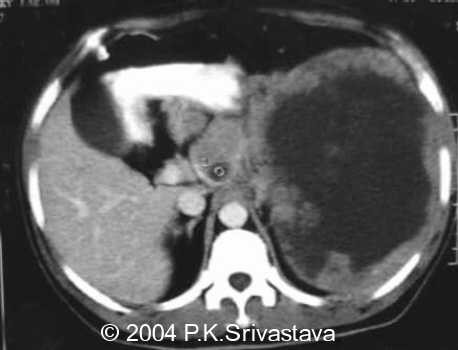
Splenic lymphoma
First published on SonoWorld
Case Presentation
A young man presented with a tender mass in the left upper quadrant. An abdominal ultrasound was performed.


Differential Diagnosis
Lymphoma, splenic abscess
Final Diagnosis
Hodgkin`s lymphoma
Discussion
Lymphomatous infiltration of the spleen is usually seen as discreet, usually hypoechoic, masses within the spleen. In this rather advanced case, the lymphomatous tissue has essentially replaced the splenic tissue creating a large and complex mass. The presence of enlarged hypoechoic nodes in the area supports the initial impression of lymphonatous infiltration of the spleen.
Follow Up
An ultrasound guided FNA was performed which confirmed the lymphomatous infiltration. The patient subsequently also developed a massive pleural effusion due to lymphomatous pleural deposits.