Intestinal Lymphoma
Case Presentation
A 81 year old male presented with asthenia and loss of weight.

First published on SonoWorld

Differential Diagnosis
Adenocarcinoma
Lymphoma
Crohn's Disease
Infectious Ileitis
Ischemia
Carcinoid
Final Diagnosis
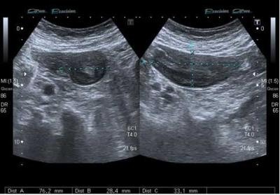
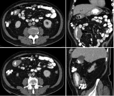
Intestinal Lymphoma (Non-Hodgkin lymphoma of the small bowel).
Discussion
Lymphoma.
"The ileum and the cecum are the most common sites of involvement by primary lymphoma in the small and large bowel, respectively. The lymphoma may also extend to the appendix.
Lymphoma of the ileocecal region most commonly manifests as single or multiple segmentalareas of circumferential thickening with homogeneous attenuation and poor enhancement. The bowel wall may display symmetric thickening and is usually markedly thickened (1.5–7 cm).
Lymphoma of the ileocecal area may also be seen as a polypoid lesion of variable size that may act as the lead point of an intussusception. Lymphoma may mimic adenocarcinoma, but in the former, the segment of bowel involved is usually longer; the transition from tumor to normal bowel is much more gradual; and associated, possibly bulky mesenteric and retroperitoneal lymph nodes are usually seen encroaching on the vessels from both sides .
A classic complication is ulceration with formation of a fistulous tract to adjacent bowel loops. An excavating mass is one that involves the entire bowel wall, with bowel entering and exiting the mass. The lumen or cavity of the mass fills with contrast material and is usually larger than the bowel entering it; this condition is referred to as aneurysmal dilatation of the bowel".
The above discussion is quoted from article:
Multi–Detector Row CT: Spectrum of Diseases Involving the Ileocecal Area
Christine Hoeffel, MD, Michel D. Crema, MD, Ahcène Belkacem, MD, Louisa Azizi, MD, Maité Lewin, MD, PhD, Lionel Arrivé, MD and Jean-Michel Tubiana, MD
doi: 10.1148/rg.265045191 September 2006 RadioGraphics, 26, 1373-1390.
URL: http://radiographics.highwire.org/content/26/5/1373.full
Follow Up
Histological appearance, immunohistochemistry and flow cytometry indicate malt lymphoma of the jejunum. (B lymphoma), with lymphomatous spread of the lymph nodes (6/6).
1. Multi–Detector Row CT: Spectrum of Diseases Involving the Ileocecal Area.
Christine Hoeffel, Michel D. Crema, Ahce`ne Belkacem,Louisa Azizi, Maite´ Lewin, Lionel Arrive´, Jean-Michel Tubiana. September 2006 RadioGraphics, 26, 1373-1390.
2. Bowel Wall Thickening on Transabdominal Sonography
Hans Peter Ledermann, Norbert Börner, Holger Strunk, Georg Bongartz, Christoph Zollikofer and Gerd Stuckmann
American Journal of Roentgenology 2000 174:1, 107-115