8. ECA vs ICA - External versus internal carotid artery

8.1 Why is it important to differentiate the internal- from the external carotid artery with ultrasound?
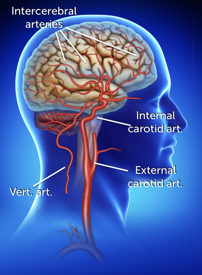
One of the most frequently asked questions, in carotid ultrasound is: “how can I tell if the vessel I am imaging is the internal- or the external carotid artery?" Especially, since the location of the vessels (and their relationship to each other) vary greatly. It can make quite a difference to the patient if a stenotic lesion or a plaque is located in the internal or external carotid. The internal carotid artery supplies the brain while the external carotid artery supplies extracranial structures of the head and neck. Therefore ischemia or an embolic event will only occur if the internal carotid artery is involved. A plaque or stenosis of the external carotid artery usually has little consequence (unless the external carotid artery provides collateral flow).
8.2 Which morphologic clues help to distinguish the internal- from the external carotid artery?
There are several observations that will help you identify the arteries. The position, size and shape are suggestive of either the internal or external carotid artery. In contrast the presence of side branches clearly denotes that the vessel is the external carotid artery.
| Morphologic clues | |
|---|---|
| Position | Large variation of the position in relationship to each other, The ICA is most commonly posterior and lateral to the ECA, When imaging the carotid artery from anterior the ECA will more frequently be closer to the transducer than the ICA |
| Size | The internal carotid artery (ICA) is more commonly larger than the external carotid artery |
| Shape | The internal carotid artery (ICA) has the bulb (the vessel is wider at its origin) |
| Branches | The external carotid artery (ECA) has side branches |


< Previous chapter: 7. Vertebral Arteries Next chapter: 9. ECA vs ICA >
8.3 How can color Doppler help to distinguish the internal from the external artery
There are several ways how both color Doppler and spectral Doppler can help to tell if the vessel you are imaging is the internal or the external artery. For example: you can use both Power Doppler and color Doppler to visualize side branches. Here are two examples.


The temporal color Doppler pattern also differs between the external and the internal carotid artery. Because the diastolic velocities are lower in the external versus the internal carotid artery we can also observe less color Doppler filling in the external carotid artery during diastole (there is more “color pulsation”).
Color Doppler also allows you to identify the internal carotid artery by detecting the area of “recirculation” of the internal carotid bulb.

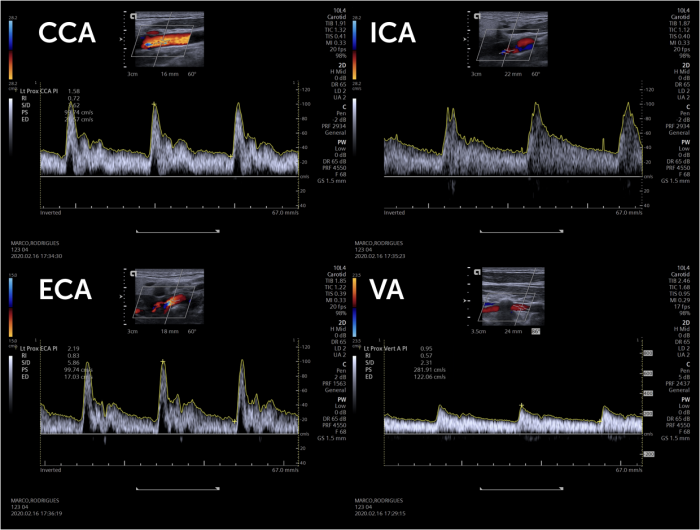
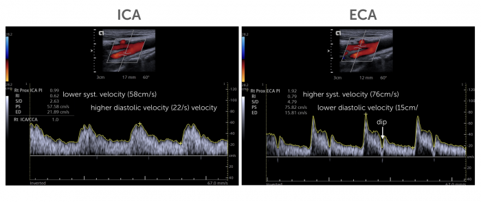
8.4 How is spectral Doppler used to differentiate between the external and internal carotid artery?
There is a distinct difference in the spectral Doppler pattern between the external and internal carotid artery. The external carotid artery supplies a “high” resistance vascular bed”, while the internal carotid artery supplies the brain which has a low resistance vascular bed.
Characteristics:
Internal carotid artery
- Higher diastolic velocities
- Often lower systolic velocities
- (Less difference between max systolic and diastolic velocities)
External carotid artery
- Initial sharp rise in velocity at systole
- Rapid decline in velocity after systole
- Sometimes retrograde flow in diastole
- Lower diastolic velocities
Note: There is a certain variation in the characteristics of the internal and external carotid artery and the patterns can sometimes look quite similar, making it difficult to differentiate the vessels. In such situations try imaging the more distal segments of the arteries. The further distal you record the Doppler signal in the internal carotid artery the higher the diastolic component will become (decrease in the S/D ratio) and the easier it will be to differentiate it from the external carotid artery.
8.5 How does the spectrum of the vertebral arteries and the common carotid artery look?
The common carotid artery supplies both a high and a low resistance bed (via the external and internal carotid artery). Therefore, the signal looks like a combination of the internal and external carotid artery. The relationship between the systolic and diastolic maximal velocities is intermediate.
The vertebral artery also supplies the brain with blood. Therefore it is a “low resistance” artery. The Spectral Doppler tracing resembles that of the internal carotid artery with a relative high diastolic velocity.
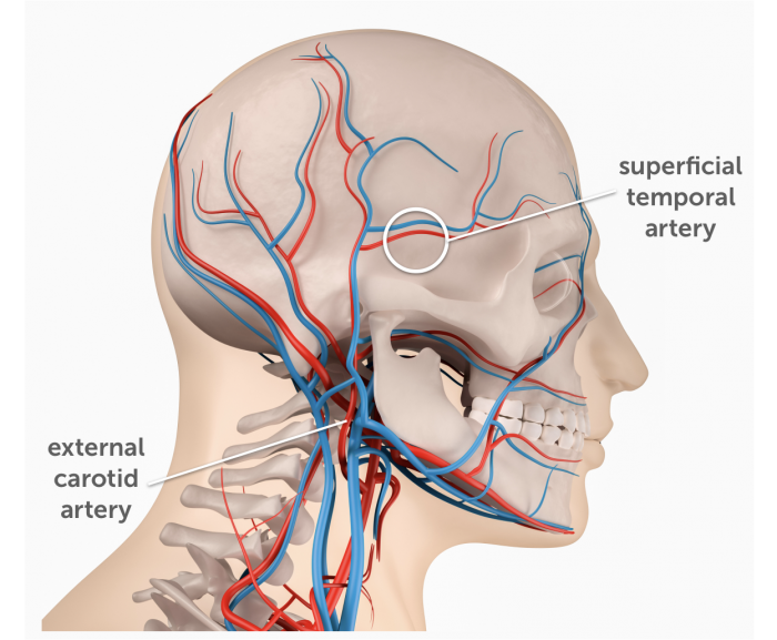
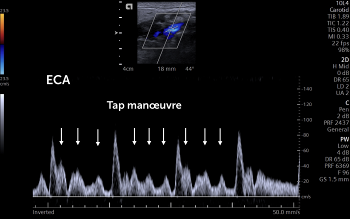
8.6 What is the “temporal tap” and how can it be used to differentiate between the internal and the external carotid artery?
The “temporal tap maneuver is used to identify the external carotid artery. Repeated compression (tapping) of the superficial temporal artery (which is located in front of the ear) causes small deflection on the spectral Doppler tracing. The maneuver is not always easy to perform. Be sure that you are really tapping the temporal artery! (you can feel the pulse of the temporal artery anterior to the ear). Perform rapid successive taps. It might be helpful to ask a colleague to perform the maneuver while you image. It is advisable to place the Doppler sample volume as far distal in the artery as possible.
Caution: The temporal tap maneuver is not always reliable as deflection waves can sometimes also be seen in the internal carotid artery.

If you like the way we teach, please leave a message!