3.3 The right ventricle
The right ventricle greatly contributes to cardiac function and is frequently affected in left heart disease. Abnormalities of the right ventricle have significant therapeutic and prognostic implications. Echocardiographic assessment of the right ventricle is more difficult than that of the left ventricle because the former has a more complex geometry, its wall is thinner, and its physiology of contraction differs from that of the left ventricle.
3.3.1 Right ventricular shape, anatomy, and function

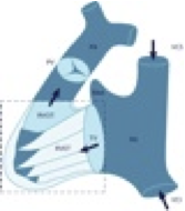
The right ventricle is difficult to describe by a simple geometric model. It has been compared to a bagpipe, but even this analogy is rather crude. The shape of the ventricle is altered in certain pathologies such as right ventricular dysplasia, volume overload, or pulmonary hypertension. In the latter you will find a "D-shaped" ventricle, which is caused by flattening of the interventricular septum. The right ventricle wraps around the left ventricle and consists of an inflow portion, the trabeculated apex, and the infundibulum. The wall of the right ventricle is much thinner (less than 5mm) than that of the right ventricle, and the inner aspect of the ventricle is marked by more numerous trabeculations, especially at the apex. Three papillary muscle groups exist: the anterior group which is the largest, the posterior, and the accessory muscles. A typical anatomical feature of the right ventricle is the moderator band. The moderator band is a muscular strand that traverses the right ventricle from the base of the anterior papillary muscle to the interventricular septum. It serves two main functions: first it conveys the right branch of the atrioventricular bundle of the conduction system and second, it prevents the right ventricle from "over-expanding". The moderator band is an important structure in echocardiography because it helps to identify the right ventricle; the latter may not be obvious, especially in complex congenital heart disease.
| Features of the right ventricle |
|---|
| Thin wall (<5mm) |
| Strongly trabeculated |
| Moderator band |
| Wraps around the LV |
Pulmonary circulation is a low-impedance, low-pressure system. The right ventricle does not have to cope with high afterload. It is therefore "weaker", smaller and thinner than the left ventricle. The right ventricle also contracts differently. The longitudinal component contributes more to contraction than the radial component. In addition, there is no rotating/twisting motion of the right ventricle.