3.5 The right atrium
The right atrium is usually the chamber that is given the least attention in echocardiography. In a way it is the right atrium (and not the right ventricle) which is the so-called forgotten chamber. Yet, the right atrium provides significant information. Enlargement of the right atrium occurs in numerous conditions, and the size of the right atrium yields prognostic information as well. Furthermore, the right atrium is probably the chamber in which benign masses and pseudotumors are most frequently found.
3.5.1 Anatomy and physiology of the right atrium
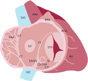
Anatomy of the right atrium, RAA = right atrial appendage, CT = crista terminalis, OF foramen ovale SVC = superior vena cava, IVC = inferior vena cava, AAo = ascending aorta, ER/EV= eustachian valve/rete CS/ThV= coronary sinus/thebesian veins, STV= septal tricuspid valve, RCA= right coronary artery, RV = right ventricle.
The right atrium is the collecting chamber for systemic venous blood returning to the heart. It functions in a very similar way as the left atrium. Right ventricular filling too is achieved via an early diastolic "passive" and a late diastolic "active" component. The right atrium is filled via the superior and inferior vena cava, the coronary sinus, and the thebesian veins. The inferior vena cava is less than 17 mm in diameter but can collapse completely during inspiration, when right atrial filling increases. The inferior vena cava is best seen from a subcostal window. There you will see it entering the right atrium shortly after it collects blood from the hepatic veins. The superior vena cava enters the right atrium from the cranial aspect, in almost the same longitudinal plane as the inferior vena cava. In a way both vena cava can be viewed as a single tube and the right atrium as a bag attached to this tube. The superior vena cava is difficult to visualize with transthoracic echo. However, in some patients it can be seen from a subcostal window.
The thebesian veins are too small to be seen with echo. The coronary sinus drains venous blood from the heart itself. It enters the right atrium from the posterior aspect, close to the atrioventricular junction, and is shaped like a funnel. It can be seen on several views, especially on the so-called coronary sinus view (see chapter: How to Image).
A prominent structure within the right atrium is the crista terminalis, which is a crescent-shaped muscular ridge between the right atrial appendage and the right atrium. It is a region in which the atrial wall is thickened (four-chamber view)
The crista terminalis of the right atrium can be confused with a mass.
The eustachian valve arises from the inferior vena cava just as it enters the right atrium. It may be quite prominent and fairly long (2 to 3 cm or more). It may lead to misinterpretation and become confused with a tumor, thrombus or even a vegetation.
The eustachian valve is important for fetal circulation - it directs blood from the inferior vena cave to the foramen ovale.
The right atrial appendage is more rectangular and "shallow" compared to the left appendage, and is less trabeculated. The right atrial appendage is not seen on transthoracic echo. However, since you will rarely see pathologies or thrombi in this region, this is no real problem.