11.4 Echocardiographic Findings in Mitral Stenosis
11.4.1 The mitral valve
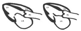
The hallmark of mitral stenosis is "doming of the anterior mitral valve leaflet". The leaflet bulges towards the left ventricle because blood is "caught" in the leaflet (similar to a sail in the wind). Doming is caused by commissural fusion and reduction of the mitral valve opening area. This is best seen on a parasternal long-axis view. The degree of doming depends not only on the severity of mitral stenosis but also on the mobility of the anterior mitral valve leaflet. When the leaflet is calcified or severely thickened and therefore "stiff", it will demonstrate mild or even no doming. Since leaflet thickening progresses with age, doming is less frequently encountered in the elderly.
The posterior leaflet typically mirrors the motion of the anterior leaflet and moves anteriorly together with the anterior leaflet during diastole - a phenomenon which can be displayed with the MMode and has previously been used as a diagnostic criterion for mitral stenosis.

| Echocharacteristics of MS |
|---|
| Doming (diastolic buldging of the AMVL) |
| Commissural fusion |
| Secondary calcification |
| Reduced valve opening |
| Leaflet tip thickening |
| Subvalvular involvement (thickend and fused tendinae) |
In rheumatic stenosis one often finds postinflammatory alterations, such as retracted and restricted leaflets, calcific deposits, or chordal rupture. Thickening is predominantly seen in the leaflet tips, but may also be present in the entire mitral valve. Very frequently one will note subvalvular involvement with thickened and fused chordae. When subvalvular involvement is very prominent, stenosis will become more "funnular".
When assessing the mitral valve it is important to use several views, especially the apical long-axis view which best displays the subvalvular apparatus, and a parasternal short-axis view at the level of the mitral valve to demonstrate commissural fusion and the opening motion of the mitral valve.
11.4.2 Other valves
Rheumatic heart disease may also affect other valves, the pericardium and the myocardium. Furthermore, the hemodynamic sequelae of mitral stenosis alter the function and morphology of the heart. Therefore, assessment of mitral stenosis should include all aspects of the disease.
There is a high prevalence of aortic valve involvement in rheumatic disease. One frequently finds mild degrees of aortic stenosis (which may progress as the patient grows older), and particularly aortic regurgitation. Thickening of the aortic valve is predominantly seen in the free edges of the cusps. Calcified deposits are observed in many cases.
The tricuspid valve is frequently incompetent, either as a result of annular dilatation or in the presence of rheumatic tricuspid valve stenosis. In latter one would find tricuspid valve doming and at least some degree of tricuspid valve inflow obstruction.

11.4.3 The heart chambers
Typically patients with isolated mitral stenosis will have a rather small left ventricle because left ventricular filling is impaired. Left ventricular function is usually normal, as the left ventricle is overloaded neither in terms of pressure nor in terms of volume. When left ventricular function is reduced, rheumatic myocardial involvement must be taken into account.
Chronic pressure overload of the left atrium leads to left atrial dilatation. However, the size of the atria not only depends on the severity of stenosis but also on the compliance of the left atrium and the presence of atrial fibrillation and/or mitral regurgitation. Further signs of elevated left atrial pressure are the presence of a dilated/expanded left atrial appendage and bulging of the interatrial septum towards the right.
The right atrium is frequently dilated as well, either due to the presence of tricuspid regurgitation, pulmonary hypertension, or both. Right ventricular enlargement may be the result of pulmonary hypertension and / or tricuspid regurgitation. Pulmonary hypertension is a frequent finding in mitral stenosis. It correlates well with the patient's symptoms and the severity of mitral stenosis.
11.4.4 Mitral stenosis and thrombus formation

Mitral stenosis predisposes to left atrial thrombus formation. The incidence of systemic embolism in untreated patients is high (10-20%) There are two main reasons why patients with mitral stenosis develop thrombi: First, blood flow within the enlarged left atrium is slow and second many patients with mitral stenosis have atrial fibrillation which leads to contractile dysfunction of the atria
| Risk of Thrombus in mitral stenosis |
|---|
| Systemic embolism occurs in 20% of all MS patients |
| 80% of with patients with thrombi have Afib |
| 45% have spontaneous left atrial contrast |
With 2D - echo you will often see spontaneous contrast in the left atria. This finding is also termed slow flow phenomena since it corresponds to the presence of slow blood flow. The presence of spontaneous contrast increases the likelihood of thrombus formation and is often seen in combination with thrombi. Most thrombi are found in the left atrial appendage. Since transthoracic echo is not ideal to visualize the appendage it is easy to miss a thrombus.
| Echocardiographic fndings in mitral stenosis |
|---|
| LA enlargement |
| Dilated pulmonic veins |
| Small LV (in the absence of LV volume overload) |
| Tricuspid regurgitation |
| Pulmonary hypertension |
| Thrombus |