Abdominal Ultrasound BachelorClass
Kidney sonography

What you will learn
A short review of the anatomic features of the kidneys and the surrounding structures will prepare you for scanning these two small organs. In this chapter we'll show you all the different approaches and scan planes for both kidneys as well as tips and tricks for difficult scanning conditions.
This chapter deals with norm variants and harmless changes of the kidneys like simple renal cysts, but also with important pathologies like renal tumors, renal stones, hydronephrosis and many more. You'll be able to detect and evaluate them after completing this chapter. Test your knowledge and solve the following case reports!
This chapter will prepare you to scan the kidneys - we'll review the anatomic features, the structures that surround the kidneys and also explain the basic image planes.
Learn how to find and scan the right kidney in longitudinal and transverse plane from different approaches.
Now let's do the same on the left side: How to scan the left kidney in two different planes.
There are lots of pathologies you can find in the kidney. Learn to detect hydronephrosis and urinary tract stones. You will see how the kidney can vary in size, form and position - we will make sure that you will always find them!
In this chapter you will learn how abdominal ultrasound can aid in the evaluation of renal tumors and how to distinguish them from cysts. What are the diagnostic criteria for harmless versus complicated renal cysts? You will find out here. Finally we will deal with the atrophic kidney disease.
Test your knowledge and see if you can solve a few cases. Here we put renal findings with ultrasound into a clinical context.
Video lectures
-
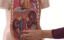 Kidneys: anatomy and How to image (basics)
This video lecture is only available for subscribers!
Get the Abdominal Ultrasound BachelorClass -
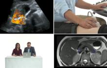 Kidneys: How to image 1
This video lecture is only available for subscribers!
Get the Abdominal Ultrasound BachelorClass -
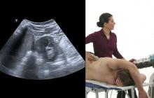 Kidneys: How to image 2
This video lecture is only available for subscribers!
Get the Abdominal Ultrasound BachelorClass -
 Kidneys: Pathologies 2
This video lecture is only available for subscribers!
Get the Abdominal Ultrasound BachelorClass -
 Kidneys: Case reports
This video lecture is only available for subscribers!
Get the Abdominal Ultrasound BachelorClass -
 Kidneys: Pathologies 1
This video lecture is only available for subscribers!
Get the Abdominal Ultrasound BachelorClass
Quiz
Start quizThis quiz is only available for subscribers!
Get the Abdominal Ultrasound BachelorClass